Home » Without Label » Left Hip Muscles Anatomy - Muscle Compartments Of The Thigh Complete Anatomy / When the hip muscles are left more or less intact, they are able to support the new.
Left Hip Muscles Anatomy - Muscle Compartments Of The Thigh Complete Anatomy / When the hip muscles are left more or less intact, they are able to support the new.
Left Hip Muscles Anatomy - Muscle Compartments Of The Thigh Complete Anatomy / When the hip muscles are left more or less intact, they are able to support the new.. I mean, the abs are the muscle. When you think of abs, what muscle do you typically think of? See anatomy hip muscles stock video clips. The posterior muscle group is made up of the muscles that extend (straighten) the thigh at the hip. They begin under the gluteus maximus behind the hipbone and attach to the tibia at the knee.
These muscles include the gluteus maximus muscle (the largest muscle in the body) and the hamstrings group, which consists of the biceps femoris, semimembranosus, and semitendinosus muscles. These are gracilis, pectineus, adductor longus, adductor brevis, adductor magnus, and adductor minimus muscles. This mri hip joint axial cross sectional anatomy tool is absolutely free to use. Pick which works for you and then. When the hip muscles are left more or less intact, they are able to support the new.
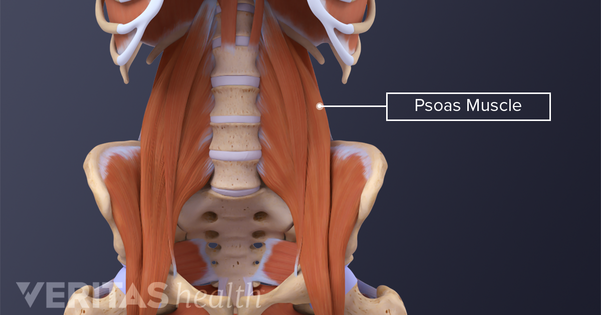
The Essential Role Of The Psoas Muscle from embed.widencdn.net Iliopsoas muscle, a hip flexor muscle that attaches to the upper thigh bone. What movements does it control? (2017, elsevier) should be consulted. One at the left hip, and one at the right hip. The anatomy of your abdominal muscles. Functionally, the hip joint enjoys a very high range of motion. The view on the left has the rectus femoris cut away to show the vastus intermedius which is below it. The hip muscles are composed of multiple flexors, extensors, adductors, abductors, and rotators that work together.
1 hip anatomy, function and common problems.
Anatomy it band pelvis muscle pelvis with muscles hip muscles muscles of pelvis tensor fascia latae psoas major anatomy pelvis tensor fascia lata pelvis muscles. Pick which works for you and then. To put it plainly, sometimes hip pain comes from the hip, but a lot of times hip pain comes from the back. The anatomy of the hip and back is comprised of numerous parts that can be injured or wear out, and many problems that occur in this area can display the exact same symptoms or pathology. This might sound like a strange question, right? The anatomy of your abdominal muscles. Patient education | concord orthopaedics. These ligaments reinforce and stabilize the hip joint(6). Use the mouse scroll wheel to move the images up and down alternatively use the tiny arrows (>>) on both side of the image to move the images.>>) on both side of the image to move the images. For detailed anatomy of pelvic bones, read anatomy of hip bone. Most modern anatomists define 17 of these muscles, although some additional muscles may sometimes be considered. But in actuality there are 4 separate muscles that contribute to your overall abdominal development. The pelvic floor muscles also help increase this pressure, which provides stability to the spine and trunk.
The anatomy of the hip and back is comprised of numerous parts that can be injured or wear out, and many problems that occur in this area can display the exact same symptoms or pathology. I mean, the abs are the muscle. The sartorius muscle is a distinctively long and thin muscle that crosses the thigh diagonally. The hip joint is a ball and socket synovial joint, formed by an articulation between the pelvic acetabulum and the head of the femur. Functionally, the hip joint enjoys a very high range of motion.
Thigh Muscle Strains Florida Orthopaedic Institute from www.floridaortho.com This mri hip joint axial cross sectional anatomy tool is absolutely free to use. The six hip adductor muscles are all located in the adductor or medial compartment of the thigh and all mainly adduct the thigh at the hip joint. Injury to the iliopsoas may cause hip pain and limited mobility. When the hip muscles are left more or less intact, they are able to support the new. Hip pain explained will teach you about the anatomy of the hips and pelvic area and how many different types of body tissues interact. They begin under the gluteus maximus behind the hipbone and attach to the tibia at the knee. The general action of these muscles is to laterally rotate the lower limb. The iliofemoral, pubofemoral, and ischiofemoral ligaments represent the thickenings of the joint capsule.
The femur may also rotate around its axis about 90 degrees at the hip.
To put it plainly, sometimes hip pain comes from the hip, but a lot of times hip pain comes from the back. It's primarily responsible for hip flexion, but it also rotates your thigh and adducts, which means it pulls your legs together when the muscles contract. If your hip flexors are too tight (or too strong) in comparison to their opposing muscles, the glutes, then your lower back muscles are likely to end up tight too — and. These ligaments reinforce and stabilize the hip joint(6). The muscles are broken down into three layers, and are primarily used to assist with the breathing process. If a strain occurs on the left side of the body, it may cause pain above the left hip. Adductor muscles on the inside of your thigh. The piriformis muscle is a key landmark in the gluteal region. Rectus femoris muscle, one of. This mri hip joint axial cross sectional anatomy tool is absolutely free to use. Hip pain explained will teach you about the anatomy of the hips and pelvic area and how many different types of body tissues interact. These muscles include the gluteus maximus muscle (the largest muscle in the body) and the hamstrings group, which consists of the biceps femoris, semimembranosus, and semitendinosus muscles. You go to the gym to train your abs.
For detailed anatomy of pelvic bones, read anatomy of hip bone. When the hip muscles are left more or less intact, they are able to support the new. These muscles include the gluteus maximus muscle (the largest muscle in the body) and the hamstrings group, which consists of the biceps femoris, semimembranosus, and semitendinosus muscles. To learn more about the lower back anatomy of the spine, please watch this video. If a strain occurs on the left side of the body, it may cause pain above the left hip.
Superficial Left And Deep Right Muscles Around The Hip Download Scientific Diagram from www.researchgate.net Hip pain explained will teach you about the anatomy of the hips and pelvic area and how many different types of body tissues interact. Adductor muscles on the inside of your thigh. They also stabilise the hip joint by 'pulling' the femoral head into the acetabulum of the pelvis. This blog post article is an overview of the muscles of the pelvis. Left hip muscles anatomy : I mean, the abs are the muscle. The hamstrings are three muscles at the back of the thigh that affect hip and knee movement. Common hip and back pain causes include injury to muscles from overuse, disc injury/degeneration, or spinal stenosis.
To put it plainly, sometimes hip pain comes from the hip, but a lot of times hip pain comes from the back.
They also stabilise the hip joint by 'pulling' the femoral head into the acetabulum of the pelvis. The hip joint is a ball and socket synovial joint, formed by an articulation between the pelvic acetabulum and the head of the femur. There are various hip flexor muscles that all work to. The pelvic floor muscles also help increase this pressure, which provides stability to the spine and trunk. What movements does it control? So can side stitches, a common and temporary athletic injury. This might sound like a strange question, right? The six hip adductor muscles are all located in the adductor or medial compartment of the thigh and all mainly adduct the thigh at the hip joint. The deep gluteal muscles are a set of smaller muscles, located underneath the gluteus minimus. It's primarily responsible for hip flexion, but it also rotates your thigh and adducts, which means it pulls your legs together when the muscles contract. Apr 13, 2020 · related posts of muscles of the lower back and hip diagram muscle anatomy gluteus. The posterior muscle group is made up of the muscles that extend (straighten) the thigh at the hip. Ligaments are soft tissue structures that connect bones to bones.a joint capsule is a watertight sac that surrounds a joint.in the hip, the joint capsule is formed by a group of three strong ligaments that connect the femoral head to the acetabulum.